dimecres, 18 de març del 2015

L:19 Observation of starch granules, micro-organism

L:19


Optical microscope (16x10 )cell of carrot 
Optical microscope (16x10) see the potato starch granules stained with IODA
Optical microscopewith oil imersio of  (100x10) see the potato starch granules stained with IODA
Opticaal microscopewith 16x10 cell of carrarot cromoplast


https://youtu.be/CMIXjBLSTFw          

video the organism cells


dimecres, 4 de març del 2015

L:17 Gram staining


L:16

 Introduction:

 In this great experiment we yogurt bacteria stained with dyes to diferend observarlos microscope after.

Objectives:

1- Differentiate yogurt bacterias.
2- Relate the staining procedure with the structure of the cells.

Materials:

- 1 Slide
- 1 Cover slip
- Tongs
- Needle
- Gram stain: Crystal violet,iodine and safranin.
- Decolorize reagents: ethanol 96%
- Microscope
- Yogur

Procedure:

Prokariotic cell observations: Gram Staining

1- Prepare a heat-fixed sample of the bacteria to be stained
2- Cover thesmear with crystal violet for an exposure of 1 min
3- Rinse with distilled water
4- Apply Iodine solution for 1 min.
5- Rinse the sample with distilled water.
6- Decolorize using ethanol.Drop by drop until the purple stop flowing.Wash immediately with distilled water.
7- Cover the sample with the safranin stain for an exposure time of 45seconds.
8- Rinse the sample with distilled water.
9- Gently dry the slide with paper(only the under pard of the slide )


Questions:

Gram Stain:

1.Complete the table thay you have below:


                  GRAM+
                   GRAM-
Crystal violet:
Color?
VIOLET
VIOLET
Iodine
Color changes
Color changes
Ethanol
Decolorize?
No Dye
Colorfast
Safranin
Color?
No Dye
Dyed Pink


Photo the yoghurt bacteria
Microscope optical

http://microbiologiabactesp3.blogspot.com.es/2009_08_01_archive.html


dilluns, 2 de març del 2015

L:16 Epidermis Cells

L:16

Introduccion:

-In this experiment we observed the microscope the epidermis of a plant which was a joint seen the wall and cellular nuclei.

Materials:

- 1 Slide
- 1 Cover Slip
- Distilled water
- Scissors
- Needle
- Join

Procedure:

Plant cells observation

1- Cut the stalk of the leek.
2- In the place of the cut,pullout the trasparent part of the epidermis
3- Using the brush, place the peel onto the slide containg a drop of tap water
4- Take a cover slip and place it gently on the peel with the aid of a needle
5- View it in the microscope.
6- Describe the change in the shape of the cells.
7- Draw a diagram with the parts of a stome: cells,guards,epidermis cells.

Salt Treatement

1- Prepare a 10% of salt solution
2- Put the salt with a dropper on the left pard of the slide .Touch the cover slip.
3- Place a piece of cellulose paper in the opposite part of the cover slip, and let the dissolution to go through your sample.


Resuls and microscope observation.


Es mostra IMG-20150302-WA0003.jpg.

100x augments
Photo epidermis cells join


Questions 

1- What is the major function of a cell membrane?   Balance of the internal environment of the external environment.

2-What is the major fuction of the cell wall? Performance featuring the contents of the cell and give rigidity, intervenes in all relations with the cell environment.

3-How does salt affect the cells shape?And the stome? Distend plant cell, putting large enough to balance the difference in osmotic pressures that occur solution external and internal pressure. The function of stomata have obrirce and tancarse as the plant that has this great remedy in case closed and tried to regulate the salt environment.


dilluns, 23 de febrer del 2015

L:15 Animals Cells VS Plants Cells

L:15

OBJECTIVE:

1- Identify the major components of cell
2- Differentiate between animals and plant cells
3- Measure dimensions of the entire cell and the nucleus.

MATERIALS:

Toothpick
2 slides
2covers slips
Watch Glasses
Forceps
Needle
Destiled Water
Dropper

Methylene blue
Iodone
Onion
Gycerine

Photo my cell:

Es mostra IMG-20150223-WA0004.jpg.


Photo the optical microscopy 600X augmentation


Photo cells onion: 


Es mostra la foto.JPG.


Photo the optical microscopy 600X augmentation


dimecres, 11 de febrer del 2015


P: 12 DNA Extraction


Introduction:

Deoxyribonucleic acid DNA is a nucleic asid that encodes the genetic instructions used in the development and functioning of all known living organisms and many viruses.
Nucleic acids compose the nucleo base( G,T,C,A ) complements G-C  A-T unid base nitrogenous
Within cells, DNA is organizet into structures called chomosomes.

Objectives:

1 Study DNA structure.
2 Understand the prosses of estracting DNA from a tissue.

Materials:

1L Erlenmeyer flask
100ml beaker
100ml graduated cylinder
Small funnel
Glass stirring rod
10ml Pipet
Knife
Safet goggles
Cheesecloh
Kiwi
Pineapple juice 1ml-5ml
Distiller water
90% Ethanol ice-cold
7ml DNA buffer
15gml dish soap
15NaCL
900ml tap water

Procedure:

1. Peel the kiwi and chop it to small piece
2.Add 8ml of buffer to the beaker
3.Mash the kiwi puree carefully for 1 minute without creating many bubbles
4. Filter the mixture
5.Add beaker contain carefull on top of the cheesecloth to fill the graduated cylinder.The juice will drain through the cheesecloth but the chucks of kiwi will not pass through into the graduated cylinder.
6. Add the pineapple juice to the green juice .This step will help us to obtain a pures solution DNA. Pineapple juice contains an enzyme thet breaks down proteins.
7. Tild the graduated cylinder and pour in an equal amount of ethanol with an automatic pipet.Put the ethanol through the graduated cylinder very carefully.You will need about equal volumes of DNA solution to ethanol
8.Place the graduated cylinted so that is eye level .Using the stirring road,collect DNA at the boundary of ethanol and Kiwi juice.Do not stir the Kiwi juice: only stir in the above ethanol layer
9.The DNA precipitatee looks like long,white and thin fibers.
10.Gently remove the stirring rod and examine what the DNA look like.


Questions:

1 What did the DNA look like?
White filaments that were precipitated in the ethanol.
2 Whty do you mash the kiwi ? Where DNA is located inside the cell?
2- To break the cells and remove the DNA that is  found in the nucleus of the cell
3 Explain what is the fuction of every compound of the buffer"soap and salt"
3- Salt is hypertonic medium that produces the break of the cell's membrane.
 Add soap extrain the proteins
4 DNA is soluble in water , but not in ethanol.What does this fact have to do with our method of extraction?
 4- When DNA comes into contact with ethanol it precipitates out of solution so you can see it forming in the clear layer above the kiwi solution. That is what I saw in the experiment


dijous, 22 de gener del 2015

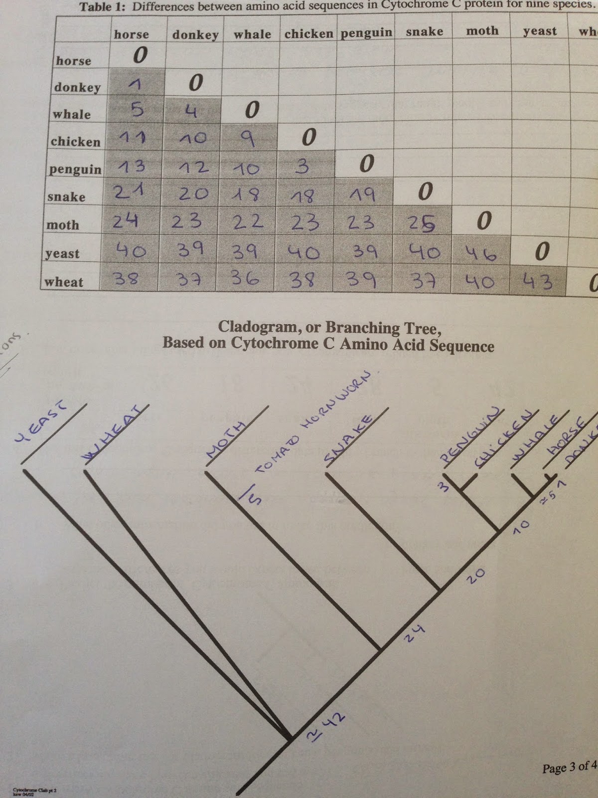
Cytochome-C Comparison Lab


Cytochome-C Comparison Lab 


PURPOSE: To compare the relatedness between organisms by examining the amino acid sequence in the protein, Cytochrome C.

Cytochome-C
Small protein from eucariotic cell,
Associated will in member of the mitocondray,
 Is molecules oxidizable ,
 Hemoproteinthe
Funcion part of electon transporchain produce energi (ATP)

This test will see the difference in cytochrome-C have the following animals

Horse -Donki- - MAMMALS
Chiken-Penguin- BIRDS
Snake-REPTILE
Silkworm moth-INSECT
Yeast-FUNGI
Wheat-PLANTS

METHOD 

Compare the amino acid sequence of Cytochrome-C in various organisms.
. Count and record the total number of differences.
Share your data with the rest of the class and complete
Make a branching tree, or cladogram with results


TABLE

Differences between amino acid sequences in Cytochrome C protein for nine species.


Horse      
Donkey
Whale
Chiken
Penguin
Snake
Moth
Yeast
Wheat
Horse  
      0


Donkey
0
     0


Whale
5
4
    0


Chiken
11
10
9
   0


Penguin
13
12
10
3
   0


Snake
21
29
18
18
19
   0


Moth
24
23
22
23
23
26
   0


Yeast
40
39
39
40
39
40
46
  0

Wheat
38
37
36
39
39
37
40
43
  0


Cladogram


QUESTIONS

1-) How many Cytochrome C amino acid sequence differences are there between chickens and turkeys? 0
2-)Make a branching tree, or cladogram for chickens, penguins, and turkeys.


3-) Predict the number of Cytochrome C amino acid sequence differences you would expect to see between
a). horse and zebra? 1-2 aprox
b). donkey and zebra?1-2 aprox


4-) What other information did you use to make this prediction?


Horse
Penguin
Snake
Tuna
Moth
Yeast
Wheat
Tomato hornworm moth
    
      26

      18

      24

     28

       5
   
      42

       38

5-) List three other things used to determine how organisms are related to each other? If they can reproduce and the offspring is fertile compering organs proce anatomic

6-) Explain why more closely related organisms have more similar Cytochrome C ? Are evolutionarily and not as separate DNA canviado not therefore less time with less canvios occur in the evolution of the species

7-) Other data, including other genes, suggests that fungi are more closely related to animals than plants.What are some reasons that the Cytochrome C data suggests that fungi, plants, and animals are equally distantly related? Based on a number of mutations as over forty are no longer compatiobles among them, have undergone many mutations